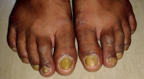
A 55-year-old male, without any comorbidity, presented to our facility with yellowish discolouration of toenails for the last 2 years. He had been diagnosed with onychomycosis by his treating dermatologist and had received multiple courses of systemic antifungal treatment without any clinical improvement. Interestingly, his past medical history was notable for some pertinent findings. A decade ago, he had undergone functional endoscopic sinus surgery for the treatment of chronic rhinosinusitis. He was hospitalised one year ago with right-sided moderate pleural effusion (exudative with lymphocyte predominance) and was conservatively managed without any sequelae. He had no symptom suggestive of any pulmonary involvement at present, or lower limb swelling, nor was he on any regular allopathic medication. Cutaneous examination revealed longitudinal ridging, nail plate thickening, transverse overcurvature and diffuse yellow discolouration involving all toenails. The cuticle and lunula could not be visualised (Figure 1). No fungal elements were detected on a potassium hydroxide mount of toenail sample. Fungal culture from the nail did not show any growth. Other mucocutaneous sites were unaffected. Systemic examination (especially pulmonary and lymphatic evaluation) and routine biochemical analysis were unremarkable. Thyroid function test and evaluations for malignancies and autoimmune diseases did not reveal any abnormality. Based on the history of pulmonary involvement, and characteristic cutaneous finding, a diagnosis of yellow nail syndrome (YNS) was established. He was treated with oral alpha-tocopherol (1,200 IU/day) and itraconazole (400 mg/day for a week every month) for three months with moderate success.
Figure 1 Yellow discolouration involving all toenails with longitudinal ridging, transverse overcurvature and nail plate thickening; the cuticle and lunula could not be visualised due to hyperkeratosis
The term YNS was described for the first time by Samman and White in 1964.1 It is a rare condition that is based on a triad associating yellow nails, pulmonary manifestations (chronic cough, bronchiectasis, pleural effusion) and lower limb lymphedema, but only two are required for diagnosis.2 Moreover, the three components are not necessarily present simultaneously, and may appear individually and sequentially, thereby making diagnosis of YNS a challenge. The complete triad is present only in 27–60% of the patients.3 Uncommon associations include cancer, autoimmune diseases and chronic sinusitis. Although aetiology remains unknown, lymphatic impairment is regularly evoked in pathogenesis; yellow nail occurs due to altered arterial circulation and Raynaud’s disease. YNS may also develop following titanium exposure from cardiac pacemakers or artificial joints.4 Oral vitamin E alone or in combination with triazole antifungals may achieve partial or total disappearance of nail discoloration.5
All three components of YNS may manifest at different times, often appearing individually. Moreover, in India, exudative pleural effusion and lymphoedema are primarily attributed to tuberculosis and filariasis, respectively. Nail discolouration of this syndrome mimics more common nail disorders like onychomycosis and chloronychia, leading to misdiagnosis. Clinicians should be acquainted with this uncommon entity.
Evaluation of lung pathology in a patient with yellow nail discoloration is of utmost importance to avoid delay in diagnosis and unnecessary treatments.