A 33-year-old female, previously well, woke up on her kitchen floor after a night out during which she drank five vodkas. She had bitten her tongue and had a mild right-sided weakness with a frontal headache. She had no significant past medical history and her only regular medication was the combined oral contraceptive pill containing ethinyloestradiol 30 mcg and levonorgestrol 150 mcg. Examination confirmed mild upper motor neurone weakness of her right arm and leg.
Following evaluation in the Emergency Department, non-contrast computed tomography (CT) head revealed hyperdensity in the interhemispheric fissure (Figure 1) and right parietal intra axial haemorrhage with mild perifocal oedema (Figure 2). The preliminary impression was that of possible subdural and intracerebral haemorrhage. However, the presence of an interhemispheric density with hyperdense cortical veins (Figure 3) suggested cerebral venous sinus thrombosis with haemorrhagic infarct, subsequently confirmed by CT venogram (Figure 4).
Figure 1 Non-contrast CT showing dense vein sign
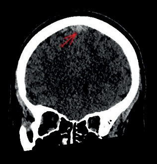
Figure 2 Non-contrast CT showing haemorrhagic infarct in right upper parietal lobe
Figure 3 Non-contrast CT showing thrombosed dense cortical veins
Figure 4 CT venogram showing triangular filling defect in the sagittal sinus, the so-called empty delta sign
Treatment was with dalteparin rather than unfractionated heparin, followed by warfarin rather than a direct acting oral anticoagulant as per the recommendations of the European Academy of Neurology.1 We also prescribed levetiracetam to prevent further seizures. When last seen at the clinic six months after discharge from hospital both her weakness and her headache had resolved completely.
Cerebral venous sinus thrombosis is a rare cause of stroke, occurring mostly in patients <50 years old and more often in women than men.2 The most common symptoms and signs are headache, seizures, focal neurological deficits and altered consciousness.3 Imaging in most patients who present in this way is likely to start with non-contrast CT, which may show appearances that bear a superficial resemblance to subdural haematoma and primary intracerebral haemorrhage, as in our case.4
The most reliable imaging modalities for diagnosis of cerebral venous sinus thrombosis are CT or MR venography.1–4 Our patient’s CT venogram showed a triangular filling defect or ‘empty delta sign’ in the superior sagittal sinus (Figure 4) confirming a diagnosis of superior sagittal sinus thrombosis. With hindsight, the hyperdensity in the interhemispheric fissure was a ‘dense vein sign’ rather than a subdural haemorrhage (Figure 1), with thrombus seen extending in the high parietal cortical veins (Figure 3) while the haemorrhage in the right upper parietal lobe was a haemorrhagic venous infarct (Figure 2).
The most likely pathogenesis in our patient was that the oral contraceptive pill caused a hypercoagulable state and that dehydration after a night out led to her venous sinus thrombosis. This in turn led to cerebral infarction and most likely precipitated a seizure. Although imaging bore a superficial resemblance to haemorrhage, the dense vein and empty delta signs were important clues to the correct diagnosis. Both appearances are recognised as important pitfalls in the diagnosis of cerebral venous sinus thrombosis.4 For the record, this patient’s presentation was unrelated to COVID-19 or to any of the COVID-19 vaccines.5